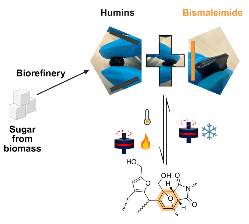
11th July 2023

SoftComp webinar programme – 1st event on 13.07.2023 at 14:00

Novel experiment opens up possibility of studying collective dynamics providing insight for better drug... (Read more)
18th October 2022

Investigating protein solution dynamics using XPCS at an XFEL

Novel experiment opens up possibility of studying collective dynamics providing insight for better drug... (Read more)
11th March 2022
Figure 2: Membrane deformations induced by actin assembly. A membrane bilayer (pink) constituting an initially spherical liposome is deformed through the growth of a branched actin network (blue marks the colour of the end of actin filaments) at its surface. Spikes (inward) and tubes (outward) are formed during this process. Note that compared to the cell geometry, the geometry here is inside out, as the ingredients of the cytoskeleton are outside the liposome. Bar 5 μm. Copyright: Cécile Sykes, CNRS

How Active Biopolymer Networks Shape the Cell Membrane

Experiments on active deformations of a cell membrane by an actin assembly may inspire new materials and approaches. Cécile Sykes from the Laboratoire de Physique de l‘Ecole Normale Supérieure reports on results raising... (Read more)
9th March 2022
Figure 1: Simulated state diagram illustrating various membrane structures for different Peclet numbers (Pe) characterizing particle propulsion strength and volume fractions (ϕ) of active particles. The three main regimes are tethering (red symbols), fluctuating (blue symbols) and bola/prolate (brown symbols) vesicle shapes. Each dot containing a grid pattern indicates the position of the nearest snapshot within the shape diagram. Simulations mimic a nearly tensionless flaccid vesicle. Copyright: authors

From Soft Active Systems to Synthetic Cells

Scientists from the ETH Zurich and Forschungszentrum Jülich highlight their recent collaborative study on a novel active system of self-propelled particles (SPPs) enclosed in a lipid vesicle, which exhibits dramatic shape changes resembling... (Read more)