How Active Biopolymer Networks Shape the Cell Membrane

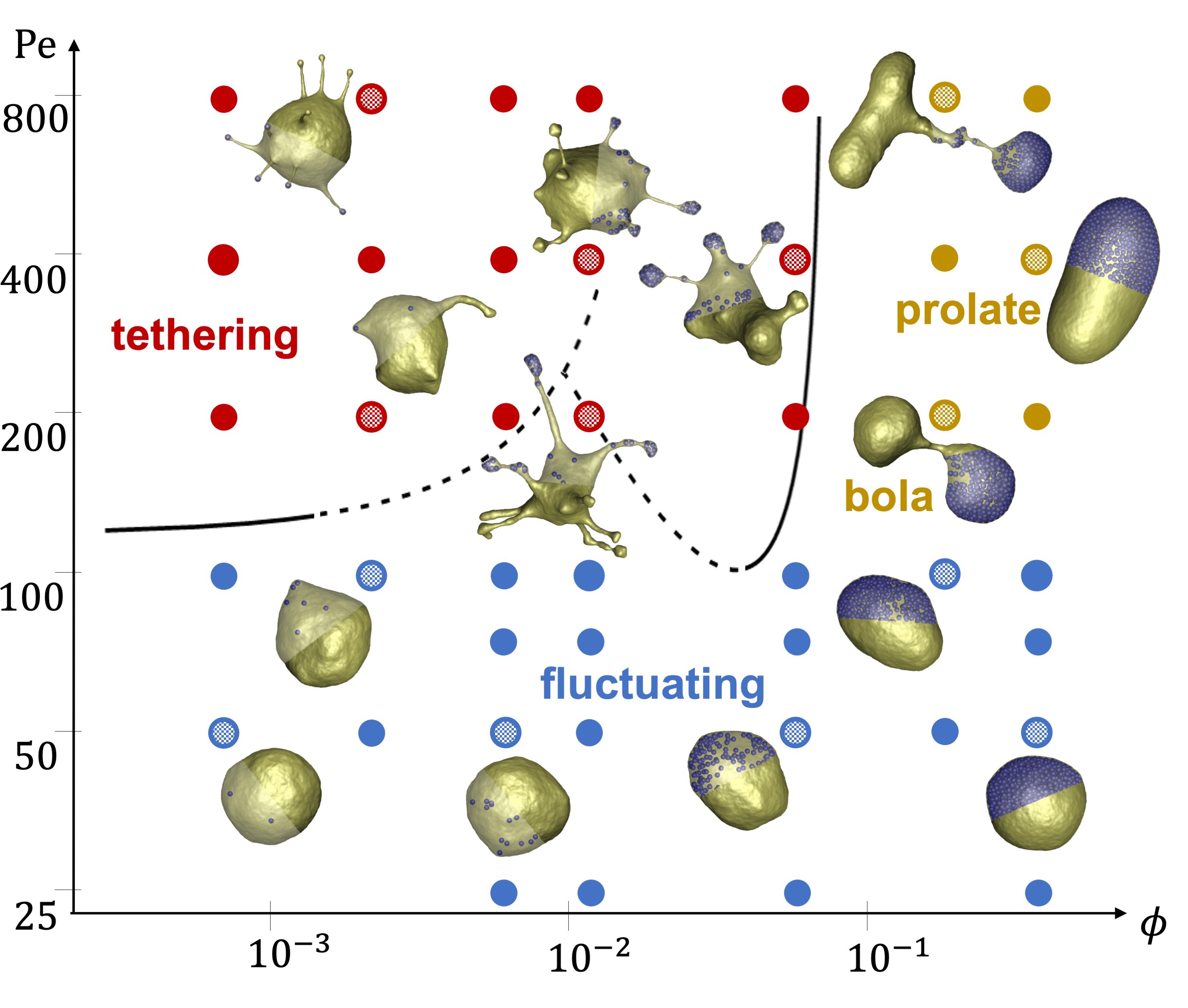
Figure 1: Simulated state diagram illustrating various membrane structures for different Peclet numbers (Pe) characterizing particle propulsion strength and volume fractions (ϕ) of active particles. The three main regimes are tethering (red symbols), fluctuating (blue symbols) and bola/prolate (brown symbols) vesicle shapes. Each dot containing a grid pattern indicates the position of the nearest snapshot within the shape diagram. Simulations mimic a nearly tensionless flaccid vesicle. Copyright: authors
From Soft Active Systems to Synthetic Cells
9th March 2022
Investigating protein solution dynamics using XPCS at an XFEL
18th October 2022

by Cécile Sykes, cecile.sykes@phys.ens.fr, Laboratoire de Physique de l‘Ecole Normale Supérieure, France

Simplistically, a living cell can be viewed as a bag filled with organelles and “cytoplasm”, a complex mixture of proteins. The bag consists of a membrane, a lipid bilayer that allows the passage of water and small molecules. Larger molecules are transported in or out of the cell by various active or passive membrane-embedded structures such as pumps and pores, or more complex channels of various conductance capacities. Cells are able to take up or release even greater volumes of material through endo- and exocytosis via the membrane.

Juxtaposed on the inner face of the cell membrane are networks of the biopolymer actin that are also present throughout the interior of the cell. This actin “cytoskeleton” ensures the dynamical and mechanical properties of the cell by assembling itself into semi-flexible filaments, i.e. with a persistence length comparable to cell size, about 10 microns. There is direct proof that the actin cytoskeleton can very robustly and reproducibly deform the cell membrane, and in particular, plays a role in the endo- and exocytosis of the cell (see, for example, references 1-4 in [1]).

Cécile Sykes, Copyright: Cécile Sykes, CNRS

Actin assembly is also known to propel intracellular bacteria such as Listeria monocytogenes, whose movement can be mimicked using beads or soft droplets/liposomes [2]. Such mimicking allows for a mechanical and biochemical understanding of the mechanism of propulsion based on squeezing and pushing forces generated by the directed growth of the actin network. Most motile events are powered by branched actin networks, formed when new actin filaments are nucleated as branches on the sides of mother filaments due to the presence of an activator at the surface of the bead, bacterium or cell membrane [3]. The force-producing actin network therefore consists of a branched, entangled, semiflexible network that constantly generates new branches at an activated surface.

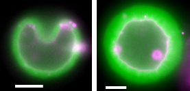
Figure 1: Buckling (left) and wrinkling (right) of a thin (left) or a thick (right) actin shell (green) grown around a membrane (pink). Bars, 5 μm. Copyright: Cécile Sykes, CNRS

Motile cells, including various mammalian cells, also use actin assembly and dynamics for their movement. Actin assembly pushes out the cell membrane at the front while actin and molecular motors, e.g. myosin, pull forward the rear by contracting the “cortex”, a thin shell of actin (a few hundred nanometers thick) that lies underneath the plasma membrane. Experiments where a cell cortex mimic is grown outside a liposome doublet allows for the quantification of the tension generated during this contraction. The Laplace law, also known as Young’s law, is used to estimate the tension increase by monitoring the change in angle between the two liposomes during acto-myosin action [4]. Strikingly, the maximal tension increase is limited to roughly 1.3-fold, in agreement with measurements in cells.

Growing a branched actin network around a liposome unambiguously reveals the elastic properties of such a network, manifested by buckling and wrinkling behaviour [5]. The experiment consists in varying the network growth time to obtain a range of actin shell thicknesses from a few hundred nanometers to several micrometers. The subsequent dilution of the external buffer generates an osmotic shock, i.e. water flows out of the liposome to equilibrate the solute on both sides of the membrane. The resulting liposome shrinkage generates a shape change of the actin shell. A buckled shape is observed for thin shells while thick shells wrinkle (Figure 1).

Such thickness-dependent behaviour is well described by a soft elastic matter model where buckling costs the least energy when the actin layer is thin, as only the edge of the buckled actin region is deformed. When the layer is too thick to buckle, wrinkling occurs involving local compression of the network. The transition between the two regimes allows for an estimate of the wrinkling wavelength (2 μm), which is larger than the predicted one (350 nm). This disparity is explained by the pre-stress in the actin shell due
to the spherical growth [5].

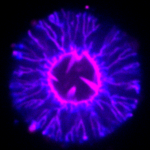
Using similar experimental set-ups with a dynamic actin assembly of branched networks on liposomes, cell-like membrane deformations are observed including tubes and spikes (Figure 2) [1]. Tubes are reminiscent of plasma membrane local shape changes during endocytosis, while spikes mimic the filopodia that cells use to investigate their surroundings. These findings indicate that cells may use the elastic properties of growing branched actin networks to deform membranes for different functions even when the biochemical components of the system are unchanged.

Figure 2: Membrane deformations induced by actin assembly. A membrane bilayer (pink) constituting an initially spherical liposome is deformed through the growth of a branched actin network (blue marks the colour of the end of actin filaments) at its surface. Spikes (inward) and tubes (outward) are formed during this process. Note that compared to the cell geometry, the geometry here is inside out, as the ingredients of the cytoskeleton are outside the liposome. Bar 5 μm. Copyright: Cécile Sykes, CNRS

Looking forward, experiments on active deformations of a membrane by an actin assembly may inspire new materials and approaches. First, the detailed mechanism of what could be called “elastic wetting” i.e. how a branched elastic network generates deformations of the substrate on which it is growing, is still to be understood. Second, the biological relevance of these findings is that deforming the membrane with cellular ingredients does not require curvature-inducing molecules as previously thought, although such molecules exist and play a role, probably in parallel with actin-only mechanisms. As often in biology, robustness is ensured by redundancy. Our results showing endocytosis-like structures with a simple set of actin-binding proteins raise the hypothesis as to whether endocytosis could be controlled for medical purposes, e.g to make it more difficult for a virus to enter cells by temporarily changing the dynamics of the actin network locally, thus altering membrane deformation to alleviate or completely prevent viral infections.

References

[1] C. Simon et al., Nature Physics 15, 602 (2019).
[2] J. Plastino and C. Sykes, Curr Opin Cell Biol 17, 62 (2005).
[3] C. Sykes and J. Plastino, Nature 464, 365 (2010).
[4] V. Caorsi et al., Soft Matter 12, 6223 (2016).
[5] R. Kusters et al., Soft Matter 15, 9647 (2019).

Research Gate
Research Gate